Home Cardiology and Pulmonology
How to Diagnose
What is Cardiopulmonary Disease | How it Affects Health | How to Use QOCA | Why QOCA
Diagnostic Methods
Diagnosis of cardiopulmonary diseases requires a comprehensive evaluation, including clinical history, physical examination, and various diagnostic tests. Here's an overview of the different tests used to diagnose cardiopulmonary diseases.
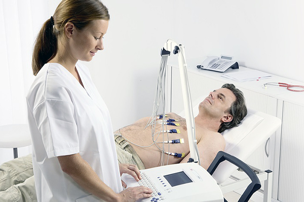
CardiacAssessment1
- Electrocardiogram (ECG or EKG). An ECG is a quick and painless test that records the electrical signals of the heart. It can identify irregular heart rhythms, which may result from various conditions, including arrhythmias, ischemia, and heart enlargement.
- Echocardiogram: This noninvasive exam uses sound waves to create detailed images of the heart in motion. It shows how blood flows through the heart and valves, helping assess the heart's structure, surrounding valves, and their function.
- Blood tests. Certain heart biomarkers, such as troponins and C-reactive proteins, gradually leak into the blood after heart damage from a heart attack. Blood tests can detect these proteins, which are associated with acute coronary syndromes. Additional tests may also assess cholesterol and blood sugar levels.
- Holter monitoring. A Holter monitor is a portable ECG device worn for 24 hours or more to record the heart's activity during daily activities. This test can detect irregular heartbeats that may not be captured during a regular ECG exam.
- Cardiac catheterization. This test can reveal blockages in the coronary arteries. A long, thin, flexible tube called a catheter is inserted into a blood vessel, usually in the groin or wrist, and guided to the heart. Dye is then injected through the catheter into the heart's arteries, making them more visible on X-ray images taken during the procedure.
- Exercise tests or stress tests. These tests often involve walking on a treadmill or riding a stationary bike while monitoring the heart. Exercise tests help reveal how the heart responds to physical activity and whether symptoms of heart disease occur during exercise. If you're unable to exercise, medication that mimics the effects of exercise on the heart may be used instead.
Credit: Arun Imaging
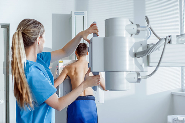
Pulmonary Assessment2
- Chest X-ray. A chest X-ray provides images of the lungs, heart, and chest wall, helping to detect pneumonia, tuberculosis, lung cancer, and other conditions.
- Bronchoscopy. A procedure that uses a bronchoscope to view the inside of the lungs and air passages, and sometimes to collect tissue samples.
- Pulmonary Function Tests (PFTs). Measure lung volume, capacity, rates of flow, and gas exchange, useful for diagnosing asthma, COPD, and other respiratory conditions.
Cardiopulmonary Assessment
- Computed tomography (CT) scan. In a CT scan, you lie on a table inside a doughnut-shaped machine. An X-ray tube inside the machine rotates around your body and collects detailed cross- sectional images of your heart and lungs to identify tumors, blood clots, or other abnormalities.1,2
- Magnetic resonance imaging (MRI) scan. A cardiac MRI uses a magnetic field and computer- generated radio waves to create detailed images of the heart and lungs, useful for detecting complex congenital heart diseases or pulmonary vascular disease.1,2
- Stethoscope. The stethoscope is an essential tool for detecting abnormal heart sounds, such as murmurs, and assessing lung sounds for issues like wheezing or crackles. It also helps monitor respiratory rates and evaluate vascular sounds, such as those in the carotid arteries. These assessments are vital for identifying signs of cardiopulmonary diseases and guiding further diagnostic testing and treatment.3,4
Credit: gorodenkoff
Advancements in medical technology have led to a growing variety of diagnostic tools, such as the ones mentioned above. While these tools can improve diagnostic accuracy, they often come with high costs and complex operations. Despite this, the stethoscope remains a widely used instrument for primary examinations, helping physicians determine the next steps in patient care.
Conventional Stethoscope
The stethoscope is a crucial tool in clinical assessments that guides further diagnostic testing and treatment.
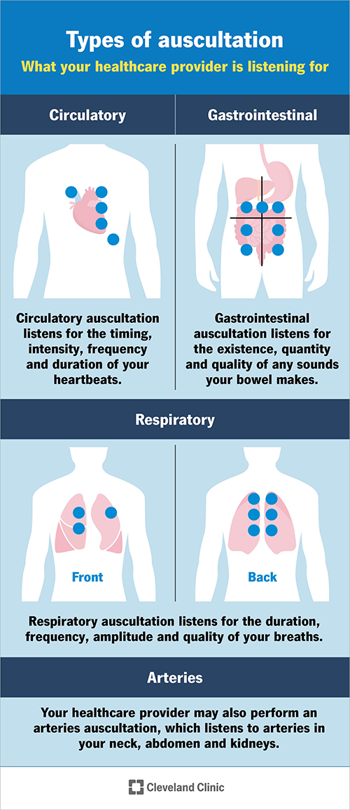
Healthcare providers commonly use a stethoscope to listen to internal sounds from the heart, lungs, arteries, and abdomen for primary examinations. Various stethoscopes are available, with many featuring two sides: the diaphragm and the bell. The diaphragm is the larger flat side and is typically used to hear high-pitched sounds, including abnormal sounds like S2 splitting in the heart, crackles in the lungs, or hypoactive sounds in the bowels that may identify blockages or paralytic ileus. The bell is the cone-shaped side and is usually used to hear low-pitched sounds, such as heart murmurs and the sound of turbulent blood flow, also known as a bruit. Bruits can indicate artery narrowing, potentially signaling a dangerous aneurysm that requires further investigation.5,6

Credit: Soup.io- Advantages and Disadvantages of a Digital Stethoscope

Credit: Bluecinema
Credit: Cleveland Clinic
Digital Stethoscope
Preserve the Past, Empower the Future: The New Era of Stethoscopes!
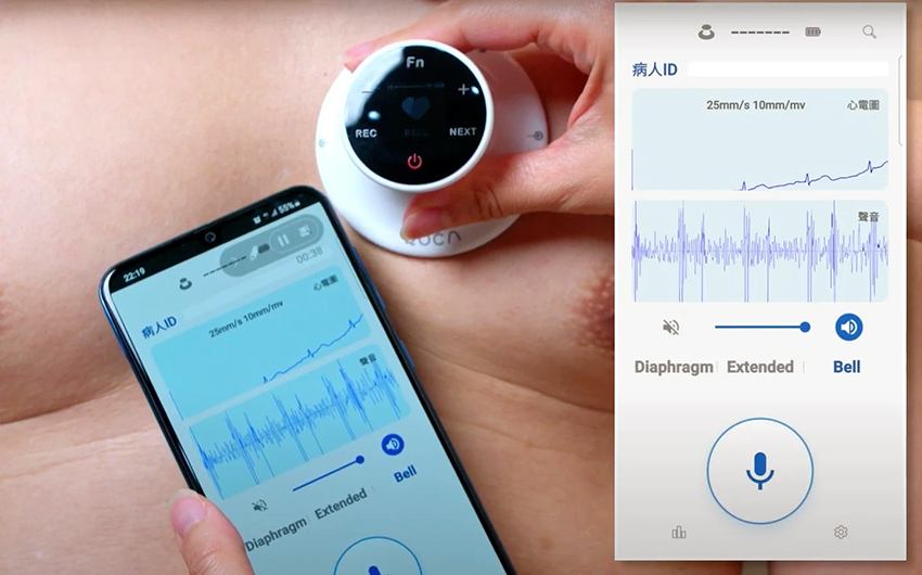
A stethoscope is a versatile tool used to detect various conditions by listening to the sounds produced by the lungs, heart, and other internal areas, such as the gastrointestinal tract. Advanced digital stethoscopes convert acoustic sounds into electronic signals, enhancing audio clarity, reducing ambient noise, and amplifying sound volume by up to 96%.7 This makes it easier for healthcare providers to detect subtle changes in sound waves, including murmurs and other nuances. Additionally, these signals can be visualized and transmitted to a computer or mobile device for AI analysis and further use.8
Modern technological advancements have greatly enhanced the development of cutting-edge medical equipment. Digital stethoscopes offer features like visualization, sound amplification for precision, and AI analysis for interpreting heart and lung sounds. Their wireless connectivity enables multiple devices to link simultaneously, promoting real-time collaboration among healthcare professionals and improving patient communication. These tools also serve as valuable resources in training, allowing medical professionals to learn from live readings.8
With these advancements, healthcare providers can continue using conventional stethoscopes while integrating advanced digital versions to improve diagnostic accuracy and patient care. By leveraging these technologies, user’s can save time, obtain precise data, clarify health issues, and deepen their understanding of patients' conditions.8
Credit: Quanta QOCA
Key Differences Between Conventional and Digital Stethoscopes8
Conventional Stethoscope
- Technology and Sound Transmission: Uses analog technology with earpieces, a diaphragm, and a bell to detect and transmit body vibrations.
- Sound Amplification and Quality: Relies on mechanical amplification, which can be influenced by external noise and the listener’s hearing ability.
- Data Recording and Analysis: Does not record or store data; information is only available during real-time auscultation.
- Integration with Technology: Functions independently without digital connectivity.
- Educational and Diagnostic Tools: Primarily used for real-time listening and diagnosis.
Digital Stethoscope
- Technology and Sound Transmission: Employs electronic components to convert sound into digital signals, providing clearer audio and the option for digital amplification and filtering.
- Sound Amplification and Quality: Offers enhanced sound quality through digital amplification and noise reduction, making it easier to hear subtle sounds.
- Data Recording and Analysis: Can record and store data for later review and analysis, often integrating with software for visual data management.
- Integration with Technology: Connects to mobile apps or cloud systems for features like sound visualization, data storage, and remote consultations.
- Educational and Diagnostic Tools: May include additional features such as ECG integration, digital auscultation analysis, and educational resources for enhanced training and diagnostics.
The digital stethoscope not only complements the traditional version but also offers numerous potential applications driven by ongoing technological advancements, such as wearable devices, continuous monitoring, telemedicine, and smart hospitals. These innovations allow for more convenient and accurate healthcare.8
Reference :
- Mayo Clinic. (2024). Heart disease.
- American Lung Association. (n.d.). Lung Procedures, Tests & Treatments.
- Shiel J., W.C. (n.d.). What Are The Four Heart Sounds? MedicineNet
- Vorvick, L. J. (2023). Breath sounds.
- Cleveland Clinic. (2024). Auscultation.
- Mauldin, A. (2025). Auscultation. Osmosis from ELSEVIER
- Stethoscope.com. (n.d.). Digital vs. Traditional Stethoscopes: A Comparative Analysis.
- Seah, J. J., Zhao, J., Wang, D. Y., Lee, H. P. (2023). Review on the Advancements of Stethoscope Types in Chest Auscultation. Diagnostics, 13(9), 1545